Frontiers Awakening Neuropsychiatric Research Into the Stria Medullaris Development of a
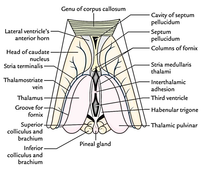
The pineal gland, habenular nuclei, and stria medullaris thalami are the principal components of the epithalamus ( Figs. 15.2, 15.4, and 15.15 ). The pineal gland consists of richly vascularized connective tissue containing glial cells and pinealocytes but no true neurons.

727 Graustufen Stria Medullaris Stockfotografie Alamy
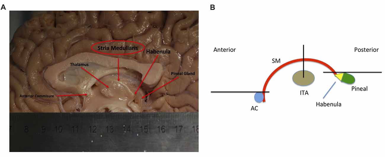
The stria medullaris (SM), (Latin, furrow and pith or marrow) is a part of the epithalamus and forms a bilateral white matter tract of the initial segment of the dorsal diencephalic conduction system (DDCS). It contains afferent fibers from the septal nuclei, lateral preoptico- hypothalamic region, and anterior thalamic nuclei to the habenula.

Image result for bed nucleus of the stria terminalis 해부학
This nucleus communicates with the rest of the limbic system via the stria medullaris thalami (along the midline of the roof of the third ventricle). Habenula 1/2. Synonyms: none. In addition to connecting the Habenular nucleus to the hypothalamus, it also connects it to nuclei of the septum (septal area).

PPT Subthalamus & Hypothalamus PowerPoint Presentation ID1159156
Die Stria medullaris thalami führt Afferenzen aus dem Riechhirn ( Substantia perforata und Regio praeoptica) zu den Habenulae. Außerdem verbindet sie die Nuclei septales, die ein Teil des Septum verum sind, mit den Habenulae. Aus der Stria terminalis lagern sich Afferenzen aus dem Corpus amygdaloideum an. An der Thalamusoberfläche folgt der.

Neurosurgery written board crash course Epithalamus part 1 habenulum YouTube
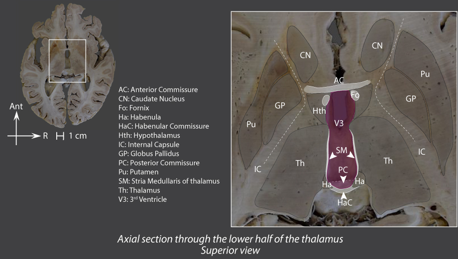
The stria medullaris, also known as stria medullaris thalami, is a fiber bundle containing efferent fibers from the septal nuclei, lateral preoptico- hypothalamic region, and anterior thalamic nuclei to the habenula. It forms a horizontal ridge on the medial surface of the thalamus . It projects to the habenular nuclei. [1]

Thalamus Anatomy, Location, Structure, Function & Physiology
Stria medullaris thalami - a bundle of fibers lying on the dorsal thalamus arising form the septal nuclei, lateral preoptic region and the anterior thalamic nuclei. References

Case MU 6664. In section 1900 the extensive demyelination of the stria... Download Scientific
It lies most medially and adjacent to the AV but is separated from AV, the stria medullaris thalami, and the ventricular surface of myelinated fibers and a glial lamella. Our results show that the.
Frontiers Awakening Neuropsychiatric Research Into the Stria Medullaris Development of a
Citation, DOI, disclosures and article data. The stria medullaris is a fiber bundle containing efferent fibers from the septal nuclei, lateral preoptico-hypothalamic region, and anterior thalamic nuclei to the habenula. It forms a horizontal ridge on the medial surface of the thalamus.
Thalamus Earth's Lab
At the time of birth (P0), the cerebral cortex is unformed, but two prominent fibre bundles are apparent in the forebrain: the medial forebrain bundle and the stria medullaris thalami.
Thalamic Nuclei Connections, Functions & Anatomy Kenhub
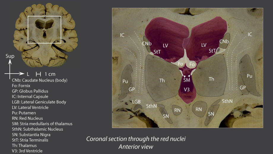
The stria medullaris thalami (SM) is the major afferent pathway to the LHb, a small nucleus . located on the dorsomedial surface of the caudal thalamus, adjacent to the third ventricle 11. The
Dorsal thalamus Braininteratlas
stria medullaris (redirected from Stria medularis thalami) stria medullaris A thin axon tract that originates in the septal nuclei, the hypothalamus, and the anterior thalamic nucleus and synapses in the habenular nuclei of the epithalamus. It is part of the limbic system. Synonym: stria medullaris thalami See: limbic system for illus
PPT Thalamus PowerPoint Presentation, free download ID9508009
Stria Medullaris. The Stria Medullaris (SM), (Latin, furrow and pith or marrow) is a part of the epithalamus and forms a bilateral white matter tract of the initial segment of the dorsal diencephalic conduction system (DDCS). It contains afferent fibers from the septal nuclei, lateral preoptico- hypothalamic region, and anterior thalamic nuclei.
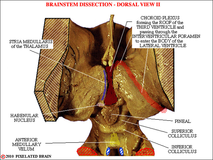
Pixelated Brain Module 2, Section 2 Dorsal views of the brainstem
It is the caudal part of the forebrain (prosencephalon) that occupies the central region of the brain. The diencephalon is comprised of the: Epithalamus Thalamus Subthalamus Metathalamus Hypothalamus In the following article, we will explore the anatomy of different parts of the diencephalon as well as their function. Contents Function

BULBUS Levent SARIKCIOLU Medulla oblongata Bulbus Myelencephalon Truncus
Stria medullaris thalami - e-Anatomy - IMAIOS Human anatomy 2 Human body Parts of human body Regions of human body Musculoskeletal systems Visceral systems Integrating systems Endocrine glands Cardiovascular system Lymphoid organs Nervous system Central nervous system Gray matter White matter Reticular formation Ependyma Meninges Brain Cerebrum

the structure of the human ear and its major parts labeled in this diagram are shown
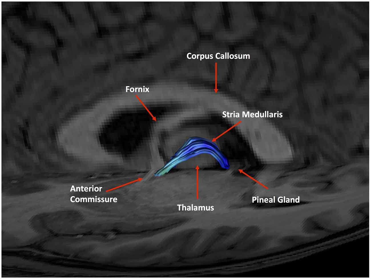
Identification of the stria medullaris thalami using diffusion tensor imaging Identification of the stria medullaris thalami using diffusion tensor imaging Neuroimage Clin. 2016 Oct 26;12:852-857. doi: 10.1016/j.nicl.2016.10.018. eCollection 2016. Authors Ryan B Kochanski 1 , Robert Dawe 2 , Daniel B Eddelman 1 , Mehmet Kocak 3 , Sepehr Sani 1
Dorsal thalamus Braininteratlas
Stria medullaris of thalamus | Anatomical Terms Pronunciation by Kenhub Anatomical Terms Pronunciation by Kenhub 8.06K subscribers Subscribe 7 Share 653 views 1 year ago Organs - how to pronounce.